New Study Solves Long-Standing Mystery of Starfish Body Structure

Bioneers | Published: February 13, 2024 Restoring Ecosystems Article
Looking at a starfish, you would not necessarily suppose it is a close relative of humans, nor that before it develops into the five-pointed creature we are familiar with, it has a body structure resembling our own.
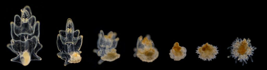
A new study published in Nature has solved a long-standing mystery for biologists about the development of starfish, or sea stars, and their body layout, revealing that the entire sea star body, including its five “arms,” is actually better described as a head. Through a process that uses genetic and molecular tools to map out their body regions, researchers were able to study how sea stars transform from a bilateral organism (the head-to-toe symmetry also found in humans) in their larval form into a creature with fivefold symmetry that is unique in the animal kingdom. One of these microscopy images is the featured image for the Bioneers 2024 Conference.
“For more than a century, biologists have been really puzzled by how this five-axis body evolved from bilateral symmetry, and how can you compare an animal with five axes like a sea star to their biological relatives, such as us,” said postdoctoral scholar Laurent Formery, the lead author on the study.
Formery said the findings not only give us new knowledge about the development and body structure of the sea star but also a window into how evolution has led to such vast differences among species.
“By conducting these types of analyses on a range of animals, you can progressively reconstruct the story of animal evolution,” he said. “That’s important because that’s also telling us where we come from.”
Though they have vastly different systems from our own, sea stars and other echinoderms — which include sea urchins, sand dollars, and sea cucumbers — are closely related to humans. A better understanding of their body plans and development can provide insight into how other animals may have evolved and arrived at their different features. The study found that the center of the sea star, as well as the center of each “arm,” has a region that actually functions like a head, with a tail-like region along the perimeter, lacking a region in between that functions like a trunk. In that way, Formery said the study found the sea star is essentially a head without a body — a notion even he admits feels disconcerting.
“When you look at a sea star, you’re basically looking at a head-like animal and it’s missing the entire trunk,” Formery said. “It seems that during evolution, this group of animals, the echinoderms, have re-engineered the anterior part of their body into a pentaradial configuration instead of developing into a head and trunk organism like most other animals do.”
To arrive at their findings, Formery and the others involved in the study used genetic and molecular tools to create a 3-D map of the sea star’s gene expression and nervous system. The mapping process creates three-dimensional renderings of the sea star’s internal structure, resulting in arresting images of the sea star’s internal structure. One of these images, which shows the nervous system of a juvenile sea star, won the 2023 Evident Global Scientific Light Microscopy Award.
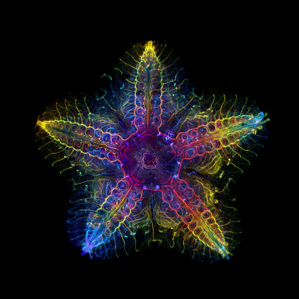
Formery said that as part of a technique called immunostaining, antibodies coupled to a fluorescent molecule are used to recognize particular proteins involved in making neurons, allowing components of the nervous system to be directly observed under a fluorescent microscope. The microscope has the ability to observe thin layers of the sea star, and by designating those layers with different colors, a 2-D image or 3-D mapping is developed. The result is a multi-layered image of the seastar’s nervous system that shows its complexity in striking rainbow hues.
Formery hopes the sea star images’ ability to grab the viewer’s attention will bring more focus to the organisms and their importance to the ocean ecosystem. For animals like sea stars, that attention is sometimes hard to come by.
“There is a strong bias in conservation efforts toward nice looking animals, so everybody cares about pandas, and nobody cares about random worms that are as endangered and as important,” he said.

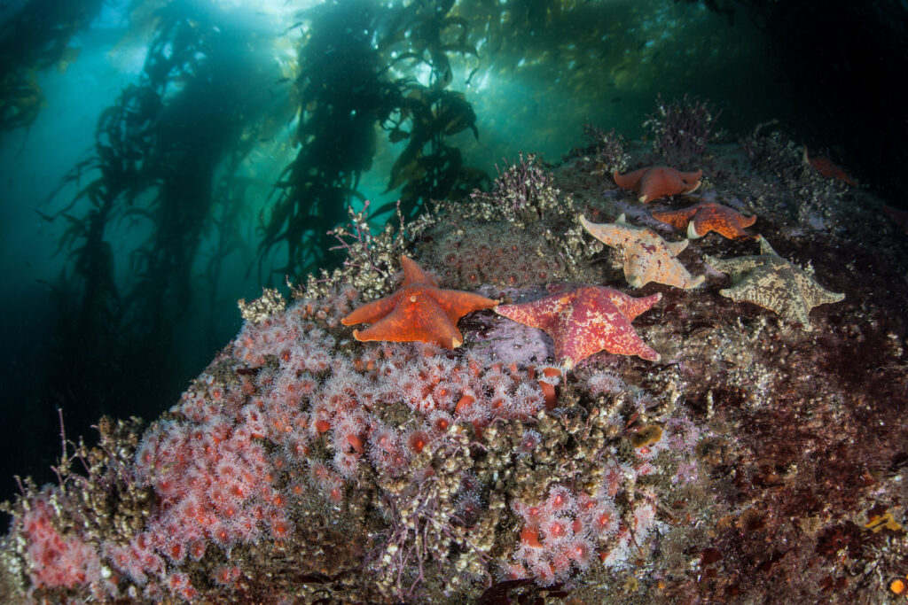
Formery emphasized that though sea stars may appear somewhat inert when observed in their habitat, this belies the crucial roles they play in their ecosystem, such as helping preserve kelp forests.
“They’re actually the predators of their environment,” Formery said. “…Sea stars prey on a lot of sea urchins and mussels, so when you remove them, it’s a big problem for the kelp forest because then the sea urchins just explode — and they eat everything.”
While the point of Formery’s microscopy work is to advance research on sea stars and other echinoderms, that beauty can sometimes result is not lost on him. By selecting particular colors to represent the various layers, he also has a hand in the visual effect. The colors, though, simply serve to illuminate the inherent beauty of the sea star.
“Sometimes you just get very beautiful samples, when the shape is perfect, undamaged, and looks exactly as you’d like it to look,” Formery said.
The study, “Molecular evidence of anteroposterior patterning in adult echinoderms,” was published in the Nov. 1, 2023, issue of “Nature.” Formery is a postdoc in the labs of Christopher Lowe at the Stanford School of Humanities and Sciences and Daniel S. Rokhsar at the University of California, Berkeley. Lowe is also a researcher at Hopkins Marine Station and senior author of the paper.
Header image credit: LiPo Ching / Stanford University